Marcel Oberlaender

Speaker of Workshop 2
Will talk about: Quantitative anatomy for assembling large scale neural networks
Marcel Oberlaender is currently working as a Senior Research Associate at the recently established Max Planck Florida Institute in Jupiter/Palm Beach, USA. Together with the inaugural director Bert Sakmann he is setting up a department for “Digital Neuroanatomy”. He will focus on quantitative morphology of individual neurons, small neural ensembles and functional networks.
Marcel Oberlaender studied Physics at the Universities of Heidelberg, Germany and Melbourne, Australia. He received his Diploma in 2006, after developing a new method for automated tracing of complete three-dimensional dendrite and axon morphologies in the department of Cell Physiology at the Max Planck Institute for Medical Research in Heidelberg, Germany.
In 2009 he received his Ph.D. in Physics for his work at the Max Planck Institute of Neurobiology in Munich, Germany, generating an anatomically realistic neural network model of a cortical column in primary somatosensory cortex of rats. He developed several methods to obtain and quantify anatomical data, such as neuron distributions, neuronal cell-types or connectivity in small circuits.
Simulating neuronal activity within network models, he is aiming to gain mechanistic understanding of network function underlying behavior or malfunction in a diseased brain.
Sensory deprivation, as well as neurodegenerative diseases, such as Alzheimer’s, cause substantial changes in brain function and anatomy, which ultimately result in behavioral deficits. Therefore, over the last 5 years, Marcel Oberlaender and his colleagues developed methods to image and quantify 3D neuron and neuronal network anatomy. These methods allow determining the number and three-dimensional distribution of all neurons in large volumes of brain tissue, the tracing of all processes from individual neurons, their classification and interconnection to realistic neural networks (see Figure).
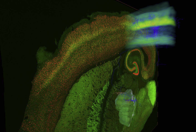
Illustration of the “Networks in silico project”. High resolution 3D image stacks of the entire brain lay the foundation to quantify the structure and 3D distribution of all neurons within functional neuronal networks. Here, the whisker-related thalamocortical pathway in rats is reconstructed.
So far these methods were limited to certain brain regions such as the somatosensory cortex or thalamus. However, recent developments in imaging techniques and computing power will allow in principle the application of these methods to the entire mouse or rat brain. The department of “Digital Neuroanatomy” at the newly founded “Max Planck Florida Institute for Integrative Biology and Neurosciences” therefore aims to determine the total number and three-dimensional distribution of all neurons in brains of “normal” mice. The resultant “cellular atlas” of the mouse brain will function as an unbiased reference for anatomical changes at cellular level caused by sensory deprivation or disease.
