Workshop 2
Chair: Mark Ellisman
Understanding the complexities of the brain is a grand challenge,
partly because of the wide range of scales involved. Many investigators
are now pushing forward to surmount the technical hurdles associated
with determining the wiring diagram of specific regions of the brain.
Modern light 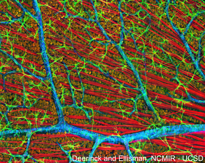 microscopic
methods do not provide adequate resolution for tracking the fine
details of cellular processes. Higher resolution imaging methods, like
3D electron microscopies, are difficult to apply to the very large
volumes of brain tissue, which are required for such mapping. However,
recent successes suggest that efforts to accelerate the development of
computer-automated methods of both light and electron microscopy may
ultimately enable more complete spatial mapping of nervous systems.
Presentations in this session will consider these issues and describe
progress in imaging cell types and their detailed relationships within
key structural domains of nervous systems. The integration of these
large and high-resolution multiscale volumes with emerging atlas
frameworks will also be discussed. This session will consist of three
lectures and a subsequent panel discussion.
microscopic
methods do not provide adequate resolution for tracking the fine
details of cellular processes. Higher resolution imaging methods, like
3D electron microscopies, are difficult to apply to the very large
volumes of brain tissue, which are required for such mapping. However,
recent successes suggest that efforts to accelerate the development of
computer-automated methods of both light and electron microscopy may
ultimately enable more complete spatial mapping of nervous systems.
Presentations in this session will consider these issues and describe
progress in imaging cell types and their detailed relationships within
key structural domains of nervous systems. The integration of these
large and high-resolution multiscale volumes with emerging atlas
frameworks will also be discussed. This session will consist of three
lectures and a subsequent panel discussion.
